Before endovascular treatment is important scales defined by levels of vascular occlusion and degree of occlusion and following the same degree of vascular recanalization and reperfusion.
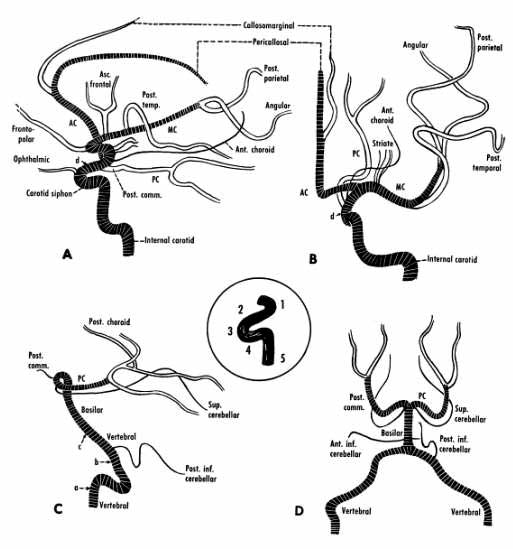
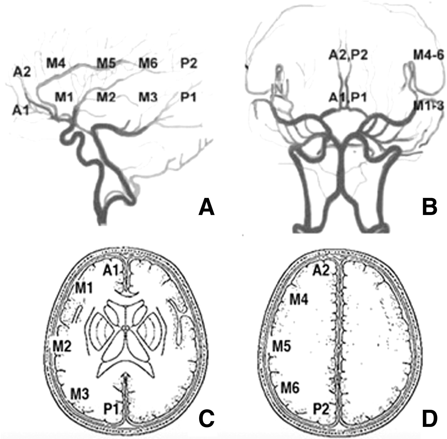
The levels are as arterial segments of the occluded artery. In ACM (M1, M2 when the artery is divided in branches Rolandic M3), ACA (A1: precommunicating artery, A2: postcommunicating anterior), ACP (P1: pre-communicating posterior, postcomun. post P2 ..), basilar (proximal third, middle or distal ) and vertebral (V1-ostium, vertebral channels:V2, V3-step intracranial and extra-intracranial V4). Carotid divide it into extra- and intracranial (in another section diagrammed).
The degree of recanalization is determined by the TIMI scale:
TIMI: 0 absence of contrast pass through the occluded vessel.
TIMI: 1 minimum or filiform pass of contrast.
TIMI: 2 passes all branches contrast unfilled.
TIMI 3 complete recanalitation.
The degree of recanalization and reperfusion was achieved with the TICI:
TICI: 0: complete occlusion. Absence of contrast passage through occluded vessel
TICI: 1: filiform passage of contrast with virtually complete distal branches.
TICI: 2: passage of contrast filling distal branches of the treated vessel. It is subdivided into:
2a: Reperfused less than two thirds of distal branches.
2b: reperfuses more than two-thirds but complete.
TICI3: complete recanalization.
TICI scale is the most useful since it not only values recanalization but also reperfusion It is the most widely used in publications and presentation of results after thrombectomy and successful (2b and 3 groups).

El punto de corte para considerar una procedimiento o resultado exitoso es cuando se consigue un nivel 2b o el 3. Un punto clave en su evaluación es discernir entre el 2b y 2a un mismo paciente. Puede variar su valoración por diferentes observadores y valoración externa dado que algunos médicos consideran los 2/3 de perfusión el límite y otros la 1/2 de ramas opacificadas y no siempre se valoran todas las proyecciones o realizan todas las proyecciones para valorar el grado de reperfusión de forma exacta.
The cutoff point for considering a procedure or successful outcome is when a level 2b or 3. A key point is achieved in their assessment is discern between 2a vs 2b in the same patient. You can obtain several assessment by different observers and external assessment. The main reason is that some doctors believe perfusion score 2b is when constrast is filling 2/3 of vascular territory and other only opacified branches in 1/2 vascular territory after vascular contrast infusion. Also not always all are projections performed to assess the degree of reperfusion exactly.


Currently the level of effective recanalization after thrombectomy 2b-3 score is around 70-85% according to current devices stent-retrievers.
Other vascular radiological data useful in assessing reperfusion therapy and its results are the collateral scales.
Check other parts of www.hubstroke.com to learn more about stroke NEWS.
.