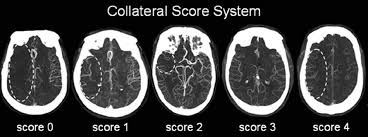
Different scales are used to assess with techniques CT / MR perfusion and therefore the viability of the tissue to initiate proceedings and the possibility of extending the time window endovascular procedures. Mainly they were initially assessed and validated with angiography but can also be evaluated with CT angiography before the start and selection of cases for a procedure.

However new softwares as RAPID (iSCHEMIA VIEW) let us to get a good evaluation of perfusion and volume map, they can aid to select patients to endovascular treatment.
Check other parts of this web address to learn more.